
Lung diseases can affect breathing, oxygen delivery, and normal lung function. Conditions such as asthma, chronic obstructive pulmonary disease (COPD), pneumonia, and pulmonary fibrosis can cause similar symptoms, including persistent cough, wheezing, and shortness of breath. Finding out what is causing these symptoms helps determine the most suitable treatment. Different Ways to Diagnose Lung Disease include physical examinations, blood tests, sputum tests, lung function tests, and imaging studies.
This article explains nine common methods used to diagnose lung diseases and why each test is important.
Quick Summary
- Medical history and physical examination: Identify respiratory symptoms, possible causes, and risk factors.
- Pulse oximetry: Estimates blood oxygen saturation (SpO₂).
- Blood tests: CBC, CRP, ESR, PCT, and ABG help assess infection, inflammation, and respiratory function.
- Sputum examination and culture: Help identify respiratory microorganisms, including tuberculosis-causing bacteria.
- Spirometry: Measures airflow and helps evaluate obstructive lung diseases such as asthma and COPD.
- Comprehensive PFTs: Assess lung volumes and gas-transfer capacity.
- Chest X-ray: Detects common lung abnormalities, infections, and fluid around the lungs.
- Chest CT scan: Provides detailed images of lung tissue, airways, and blood vessels.
- Bronchoscopy and lung biopsy: Allow direct airway examination and collection of respiratory samples or tissue.
Important Note: There is no one test that diagnoses all lung diseases. Tests used will depend upon the symptoms, medical history, physical examination, and suspected diagnosis.
Importance of Lung Disease Diagnostic Tests
Diagnostic tests help doctors find the actual cause of breathing problems, detect lung abnormalities, and check how well the lungs are working. They are useful for:
- Detecting possible respiratory conditions, including asthma, COPD, and pulmonary fibrosis.
- Assessing airflow, lung capacity, and oxygen exchange.
- Identifying respiratory infections and inflammation.
- Detecting structural abnormalities in the lungs and airways.
- Evaluating disease severity and monitoring changes over time.
9 Different Ways to Diagnose Lung Disease
1. Medical History and Physical Examination
Medical history and physical examination are initial methods used in the diagnosis of suspected lung disorders. These initial tools can indicate the probable cause of the problem and investigations required for making an accurate diagnosis.
Medical History: Doctors usually ask about the type, duration, and severity of symptoms, such as:
- Persistent cough or phlegm
- Difficulty breathing or wheezing
- Chest tightness and pain
- Hemoptysis
- Fever and frequent respiratory tract infections
- Unintentional weight loss
Doctors may also ask about smoking, workplace exposure, air pollution, allergies, previous tuberculosis, and family history of lung diseases. For instance, episodic wheezing is an indication of asthma, whereas chronic cough in smokers is indicative of COPD and bronchiectasis.
Physical Examination: During the examination, doctors check the breathing rate, chest movement, breathing effort, and oxygen levels. Breathing sounds are also listened to using a stethoscope. The commonly observed abnormal findings include:
- Wheezes: This is an indication of narrow air passages, commonly associated with asthma and chronic obstructive pulmonary disease.
- Crackles: These may be observed in patients suffering from pneumonia, pulmonary fibrosis, or pulmonary edema.
- Decreased breath sounds: These are indications of decreased air flow, pleural effusions, or pneumothorax.
- Stridor: An indication of upper airway obstruction or constriction.
Clinical Significance
Medical history and physical examination can aid in making a clinical diagnosis at first. Additional tests, such as blood tests and imaging scans, can provide more information to support the diagnosis.
2. Pulse Oximetry Test
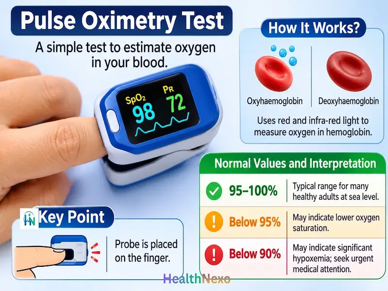
Pulse oximetry is a simple test that uses a fingertip sensor to estimate the oxygen level in the blood without drawing a blood sample. The test results are referred to as peripheral oxygen saturation (SpO₂).
Scientific Principle
Oximetry uses red and infra-red light. Oxyhemoglobin and deoxyhaemoglobin will absorb different amounts of these wavelengths. By analyzing the amount of light absorbed during pulsatile blood flow, SpO₂ can be calculated. The probe is generally placed on the patient’s finger.
Normal Values and Interpretation
- 95–100%: Typical range for many healthy adults at sea level.
- Below 95%: May indicate lower-than-expected oxygen saturation and require clinical assessment.
- Below 90%: May indicate significant hypoxemia and requires urgent medical evaluation.
These are general reference values, not universal diagnostic thresholds. Interpretation may vary with altitude, underlying medical conditions, and prescribed oxygen targets.
Clinical significance
Use of pulse oximetry can help assess oxygen levels in patients with various lung diseases including pneumonia, COPD, severe asthma, pulmonary fibrosis and respiratory failure. Low pulse oximetry results suggest hypoxia; however, they do not specify the cause of the hypoxia.
Limitations
Pulse oximetry readings may be affected by poor circulation, cold fingers, movement, nail polish, skin pigmentation, smoking, and incorrect probe placement. Pulse oximetry measures oxygen saturation but cannot detect CO₂ retention or changes in blood pH. Therefore, normal SpO₂ does not always rule out respiratory problems. Results should be interpreted alongside symptoms and clinical findings (FDA).
3. Blood Tests for Lung Diseases
Blood tests are useful in assessing infections, inflammatory responses, allergies, and breathing gases. However, they help in the diagnosis of lung diseases, but not in determining the type of disease.
1. Complete Blood Count (CBC)
The CBC test is performed to measure different parts of the blood, such as red blood cells, white blood cells, hemoglobin, hematocrit, and platelets. It also provides a count of different types of white blood cells.
Standard adult reference intervals:
- WBC: 4.0-11.0 × 10⁹/L
- Hemoglobin: male 13.5–17.5 g/dL; female 12.0–16.0 g/dL
- Platelets: 150-450 × 10⁹/L
- Absolute eosinophils: approximately 0–500 cells/µL
The reference intervals may differ depending on the lab.
Clinical significance: Elevated WBC or neutrophils could indicate the presence of an infection or inflammation. Elevated eosinophils could indicate an allergic reaction and some asthma phenotypes. Hemoglobin deficiency reduces the capacity of the blood to transport oxygen. The CBC test results cannot independently diagnose a respiratory disease.
2. C-Reactive Protein (CRP)
CRP is an acute-phase protein that is produced mainly by the liver. Its levels increase during inflammation, mainly in response to a signal called interleukin-6 (IL-6). CRP levels are usually below 5 mg/L, but the normal range may vary between laboratories.
Clinical significance: CRP levels can rise in cases of bacterial pneumonia, certain viral respiratory infections, tuberculosis, and various other inflammatory diseases. However, CRP cannot identify the specific microorganism responsible for an infection.
3. Erythrocyte Sedimentation Rate (ESR)
ESR is the measurement of the rate at which red blood cells move down the bottom of an upright tube in one hour. During inflammation, proteins such as fibrinogen cause red blood cells to stick together and settle faster.
Approximate adult reference intervals:
- Men under 50: 0–15 mm/hour
- Women under 50: 0–20 mm/hour
- Men over 50: 0–20 mm/hour
- Women over 50: 0–30 mm/hour
ESR levels may increase in people with tuberculosis, respiratory infections, and other inflammatory conditions. However, ESR can increase in many conditions, so it cannot identify or confirm a specific lung disease.
4. Procalcitonin (PCT)
Procalcitonin is the prohormone of calcitonin. Its level could rise due to infections of the body by bacteria.
Commonly Used PCT Interpretation Thresholds
- Below 0.1 ng/mL: Low concentration
- 0.1–0.25 ng/mL: Relatively low concentration
- 0.25–0.5 ng/mL: Increased suspicion of bacterial infection in some clinical settings
- Above 0.5 ng/mL: May raise greater concern for systemic bacterial infection
Important: The PCT cutoff values differ according to the clinical situation and the test used. While useful for clinical evaluation purposes, they are not absolute diagnostic cutoff values.
Clinical Significance: PCT can be used in diagnosing bacterial respiratory infections, especially for hospitalized patients. However, a low PCT level does not always mean that bacterial pneumonia is absent. Doctors should consider other clinical findings before deciding whether antibiotics are needed.
5. Arterial Blood Gas (ABG)
Arterial Blood Gases tests determine the levels of oxygen and carbon dioxide in the bloodstream. This test is performed by measuring the blood pH. This blood test is usually taken from the radial artery located in the wrist area.
Common adult reference values:
- pH: 7.35–7.45
- PaO₂: 80–100 mmHg
- PaCO₂: 35–45 mmHg
- HCO₃⁻: 22–26 mmol/L
- SaO₂: Approximately 95–100%
Values may vary with age, altitude, oxygen therapy, and clinical circumstances.
Clinical significance:
- PaO₂: Measures the partial pressure of dissolved oxygen in arterial blood.
- PaCO₂: Reflects ventilation and carbon dioxide removal.
- pH: Indicates the blood’s acid-base status.
- HCO₃⁻: Helps assess the metabolic component of acid-base balance.
- SaO₂: Represents arterial hemoglobin oxygen saturation.
Low PaO₂ implies poor oxygenation, and high PaCO₂ implies poor ventilation. ABG becomes more relevant in cases of respiratory failure, severe exacerbations of COPD, and acute respiratory disease. Contrary to pulse oximetry, ABG gives information on oxygen, carbon dioxide, and acid-base status.
4. Sputum Examination and Culture
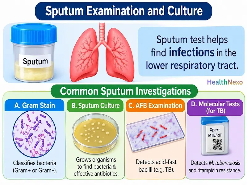
The sputum test is useful in identifying infections within the lower respiratory tract. The sputum refers to the thick mucus secreted from the lungs or airways. It may contain inflammatory cells, microorganisms, and dead cell materials.
Common Sputum Investigations
- Sputum Gram Stain: Gram staining is a microscopic procedure for classifying bacteria as being either Gram-positive or Gram-negative. It gives initial insight into the morphology of the bacteria and other inflammatory cells.
- Sputum Culture: Sputum culture involves growing microorganisms from a sputum sample in the laboratory. This test helps identify bacteria and determine which antibiotics may be effective.
- Acid-Fast Bacilli (AFB) Examination: AFB staining makes use of specific stains to identify acid fast organisms, such as Mycobacterium tuberculosis. Nevertheless, a negative AFB smear cannot rule out the possibility of tuberculosis.
- Molecular Tests for Tuberculosis: The Xpert MTB/RIF and Xpert MTB/RIF Ultra tests are molecular tests that detect the genetic material of M. tuberculosis. In addition, they also detect genetic mutations causing resistance to rifampicin. The WHO recommends the use of rapid molecular tests for the initial diagnosis of tuberculosis in many patients.
Clinical Significance and Limitations
Sputum analysis is a useful diagnostic tool for pulmonary TB, bacterial pneumonia, some fungi, and other lower respiratory tract infections. The specimen needs to be of good quality and collected from the lower respiratory tract and not mostly saliva. Contamination of the mouth, past antibiotic treatment, low number of microorganisms, and poor specimen collection can contaminate the actual results.
5. Spirometry Test
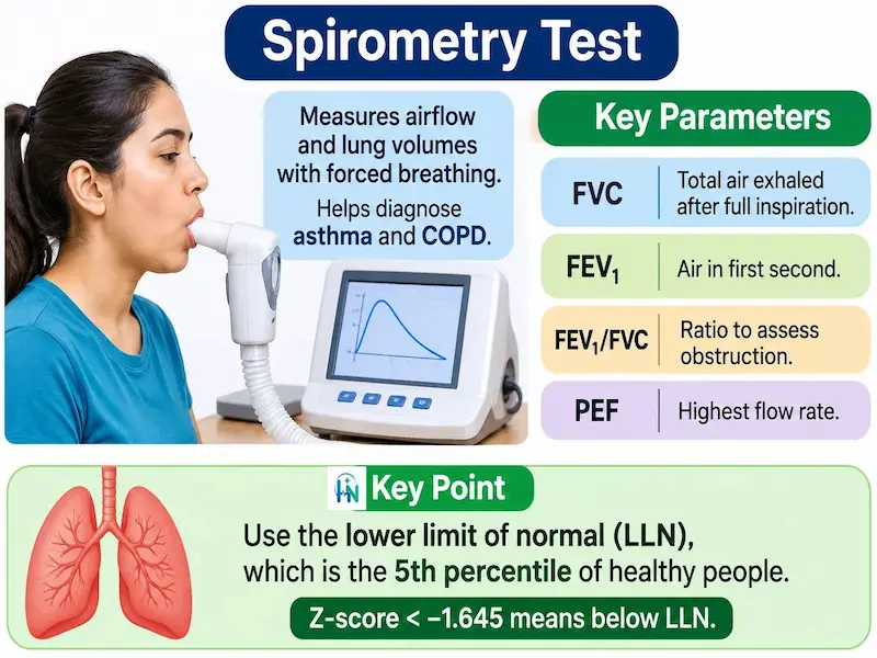
Spirometry is a crucial lung function test that measures lung volume and airflow with forced breathing. This is helpful for diagnosing obstructive airway diseases like asthma and COPD.
Scientific Principle
Air travels from the lungs to the airways during forced expiration. When there is narrowing of the airways, the expiratory airflow becomes reduced. The spirometer is used to measure airflow and volumes at different times.
Important Spirometry Parameters
- FVC (Forced Vital Capacity): Total air forcibly exhaled after a full inspiration.
- FEV₁: Volume of air exhaled during the first second.
- FEV₁/FVC: Ratio of FEV₁ to FVC, used to assess airflow obstruction.
- PEF (Peak Expiratory Flow): Highest airflow rate achieved during forced expiration.
Normal Values and Interpretation
Normal spirometry values vary according to a person’s age, sex, height, and the reference standards used. The modern approach relies on using the lower limit of normal (LLN), which corresponds to the fifth percentile of healthy individuals. A z-score less than −1.645 implies being below the LLN.
Key findings include:
- Reduced FEV₁/FVC: Suggests airflow obstruction.
- Reduced FVC with a preserved or increased FEV₁/FVC: May suggest a restrictive pattern.
- Reduced TLC: Confirms restrictive ventilatory impairment when below the LLN.
Spirometry alone cannot confirm restriction; total lung capacity (TLC) measurement is required.
COPD Diagnosis
According to the Global Initiative for Chronic Obstructive Lung Disease (GOLD), a post-bronchodilator FEV₁/FVC ratio below 0.70 indicates persistent airflow limitation. This finding is required to confirm COPD in patients with symptoms of the disease. Doctors should also consider the patient’s symptoms and medical history when making the diagnosis.
Bronchodilator Response
Spirometry may be repeated after giving the patient a bronchodilator (a medicine that helps open the airways). According to the 2022 ERS/AS criteria, an increase of more than 10% in predicted FEV₁/FVC value indicates that there is a positive response to the drug.
Clinical Significance
Spirometry helps diagnose asthma, COPD, and other conditions that cause airway narrowing. This test is also effective in the evaluation of unknown respiratory problems.
6. Comprehensive Pulmonary Function Tests (PFTs)
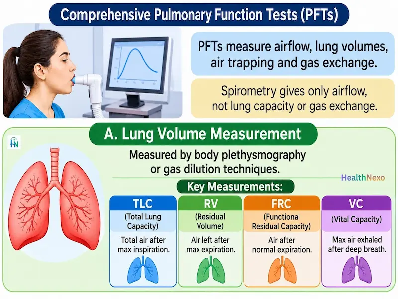
Comprehensive pulmonary function tests (PFTs) measure airflow, lung volumes, air trapping, and how well gases move from the lungs into the blood. Spirometry does not give any further information regarding the capacity of the lungs and the efficiency of gas exchange.
1. Lung Volume Measurement: Lung volumes are measured using body plethysmography or gas dilution techniques. Important measurements include:
- Total Lung Capacity (TLC): Total air in the lungs after maximal inspiration.
- Residual Volume (RV): Air remaining after maximal expiration.
- Functional Residual Capacity (FRC): Air remains after normal expiration.
- Vital Capacity (VC): Maximum air exhaled after maximal inspiration.
Clinical significance:
- Reduced TLC may confirm restrictive ventilatory impairment when below the LLN.
- Increased RV or RV/TLC may indicate air trapping, commonly seen in obstructive conditions such as COPD.
- Reduced VC may occur in both restrictive and obstructive disorders.
2. Diffusing Capacity (DLCO)
The DLCO test measures how well gas (carbon monoxide) moves from the air sacs (alveoli) in the lungs into the blood. This process depends on the size of the air sacs, the thickness of the lung membrane, blood flow in the lungs, and hemoglobin levels.
A low DLCO level may be seen in conditions such as pulmonary fibrosis, emphysema, pulmonary vascular disease, and some types of anemia.
3. Bronchodilator Response Testing
This test is performed to compare the measurements of airflow both before and after taking a bronchodilator. A change in the level of FEV1 or FVC suggests a change in airflow restriction, which can help diagnose asthma and other obstructive airway diseases.
Clinical Significance of Complete PFTs
Complete PFTs are useful for diagnosing physiological problems:
- Low FEV1/FVC: Implies airflow obstruction.
- Low TLC: Implies restrictive ventilatory defect.
- High RV/TLC: Implies air trapping.
- Low DLCO: Implies gas-transfer problem.
The PFT findings should be interpreted along with the patient’s symptoms and other clinical findings. PFT results show lung function abnormalities but not all diseases.
7. Chest X-ray
A chest X-ray produces images of the lungs, heart, airways, and other structures inside the chest. A chest X-ray uses a small amount of radiation to produce images of the lungs. It is often one of the first tests doctors recommend for a persistent cough, fever, chest pain, or difficulty breathing.
Scientific Principle
X-rays pass through the body and create images that show different structures inside the chest. Air-filled lungs appear as dark shadows, bones as white, and soft tissue/fillings as gray.
Common X-ray findings and their clinical significance
- Consolidation: It can be associated with pneumonia.
- Hyperinflation: It may result from COPD or emphysema.
- Interstitial patterns: They can suggest interstitial lung disease.
- Lung nodules and masses: They need to be investigated further.
- Pleural effusion: This means there is excess fluid surrounding the lungs.
- Pneumothorax: A condition in which air collects in the pleural space, the area between the lungs and chest wall.
- Cardiac enlargement: It results in an enlarged cardiac silhouette.
The chest x-ray is useful for evaluating pneumonia, pulmonary tuberculosis, COPD changes, pulmonary edema, pleural effusion, pneumothorax, and lung lesions.
Limitations
Small nodules, early interstitial involvement, and infections can be missed in chest X-rays. A negative X-ray does not rule out conditions like asthma. Further investigation might be needed using techniques such as CT scans, microbiological tests, or pulmonary function tests.
8. CT Scan of the Chest
Computed Tomography (CT) gives cross-sectional pictures of the lungs and the surrounding regions. It provides more detailed images of the lungs, airways, blood vessels, and lymph nodes than a regular chest X-ray.
Scientific Principle
A CT scanner uses rotating X-ray beams and detectors to collect images from different angles. A computer processes these measurements to produce detailed cross-sectional images.
Common Types of Chest CT
- Non-contrast CT: Performed without intravenous contrast.
- Contrast-enhanced CT: Uses an iodine-based contrast agent to assess blood vessels and other chest abnormalities.
- High-Resolution CT (HRCT): Provides detailed images of lung tissue and interstitial abnormalities.
- CT Pulmonary Angiography (CTPA): Evaluates pulmonary arteries, particularly when pulmonary embolism is suspected.
Common Findings and Clinical Significance
- Ground-glass opacity: May occur in infection, inflammation, or interstitial lung disease.
- Pulmonary nodules: May require follow-up or further investigation.
- Consolidation: May indicate infection or other alveolar disease.
- Emphysematous changes: May support the evaluation of emphysema.
- Interstitial thickening: May suggest pulmonary fibrosis or other interstitial abnormalities.
- Bronchiectasis: Indicates abnormal widening of the airways.
- Pulmonary arterial filling defects: May indicate pulmonary embolism on appropriate contrast-enhanced imaging.
Chest CT is helpful in assessing the condition of patients with lung cancer, lung nodules, lung fibrosis, bronchiectasis, complicated pneumonia, and emphysema. CTPA helps detect blood clots in the lungs when pulmonary embolism is suspected.
Limitations
Although CT scans provide detailed images of lung structures, they do not show how well the lungs are working. The limitations of CT include radiation exposure, allergic reaction to contrast, and additional testing of incidental findings.
9. Bronchoscopy and Lung Biopsy
Bronchoscopy is a procedure that allows doctors to look inside the airways using a thin, flexible tube with a light and camera. The procedure is also useful in collecting fluid or tissue samples for further analysis in laboratories.
Scientific Principle
During flexible bronchoscopy, a narrow flexible tube is carefully introduced through either the nose or mouth to the respiratory tract. Trachea and the larger airways can be inspected directly and samples obtained as well. Some common procedures include bronchoalveolar lavage (BAL), bronchial washing, bronchial brushing, endobronchial biopsy, and transbronchial lung biopsy.
Common Sampling Procedures
- Bronchoalveolar Lavage (BAL): A small amount of sterile saline is put into a part of the lung and then collected for laboratory testing. The sample may contain inflammatory cells, microorganisms, and other cellular materials.
- Bronchial Biopsy: Small tissue samples are collected from the airway lining and examined for inflammation, infection, abnormal cellular changes, or malignancy.
- Transbronchial Lung Biopsy: Small lung tissue samples are collected through the bronchoscope for histopathological examination.
Clinical Significance
Bronchoscopy can help diagnose lung cancer, pulmonary tuberculosis, fungal and bacterial infections, and some lung diseases. It can also help identify unexplained lung abnormalities and bleeding in the respiratory tract.
Limitations and Possible Risks: Bronchoscopy is not necessary for every patient with suspected lung disease. Possible complications include:
- Temporary coughing or throat irritation
- Bleeding, particularly after biopsy
- Infection
- Low oxygen levels during the procedure
- Pneumothorax, particularly after certain transbronchial biopsies
- Reactions to sedative medications
Bronchoscopy may be needed depending on the suspected disease, the need for samples, and the patient’s overall health.
Diagnostic Test Selection for Lung Diseases
Doctors select diagnostic tests based on the patient’s symptoms, medical history, physical examination, and suspected condition.
- Low oxygen saturation: Pulse oximetry and ABG.
- Suspected respiratory infection: CBC, CRP, sputum examination, and chest X-ray.
- Suspected asthma or COPD: Spirometry.
- Possible restrictive lung disease: Comprehensive PFTs and HRCT.
- Suspected pulmonary tuberculosis: Sputum AFB examination and molecular tests.
- Unexplained lung abnormalities: Chest CT, bronchoscopy, or lung biopsy when indicated.
For example, people with a persistent cough and fever may need a chest X-ray and sputum tests. If COPD is suspected, spirometry can help check for narrowed airways.
Conclusion
Different Ways to Diagnose Lung Disease involve a combination of clinical assessment and appropriate tests. Blood tests, sputum analysis, lung function tests, and imaging studies provide useful information about lung health. Choosing the right tests based on symptoms and clinical findings helps identify the underlying condition and guide appropriate treatment. Along with proper diagnosis and medical care, healthy daily habits and simple home remedies may help support lung health and promote easier breathing.
Frequently Asked Questions (FAQs)
Q1. What is the most common test used to diagnose lung disease?
Spirometry is a commonly used test for evaluating lung function and identifying obstructive conditions such as asthma and COPD.
Q2. Can a CBC test detect lung disease?
No. A CBC cannot independently diagnose lung disease, but changes in white blood cells, eosinophils, and hemoglobin may provide clues about infection, inflammation, allergic responses, and oxygen-carrying capacity.
Q3. Which test measures’ lung capacity?
Comprehensive pulmonary function tests (PFTs) measure lung volumes, including total lung capacity (TLC), residual volume (RV), and functional residual capacity (FRC).
Q4. Can a chest X-ray detect all lung diseases?
No. Chest X-rays can identify many structural abnormalities but may miss early disease, small nodules, and functional airway disorders such as asthma.
Q5. What is the difference between spirometry and pulmonary function tests?
Spirometry measures airflow and forced expiratory volumes, while comprehensive PFTs also assess lung volumes and gas-transfer efficiency.
Q6. Which test confirms COPD?
Post-bronchodilator spirometry showing an FEV₁/FVC ratio below 0.70 supports COPD diagnosis in patients with compatible symptoms and relevant risk factors.
Q7. Which blood test indicates lung infection?
CBC, CRP, ESR, and procalcitonin may provide supportive evidence of infection or inflammation. Sputum testing, molecular tests, and imaging may also be needed to identify the underlying infection.
Medical Disclaimer
This article is intended for educational purposes only and does not replace professional medical advice, diagnosis, or treatment. Respiratory test results should be interpreted by qualified healthcare professionals alongside symptoms, medical history, and other clinical findings.
References
- National Heart, Lung, and Blood Institute (NHLBI). Tests for Lung Disease. National Institutes of Health. Read reference
- U.S. Food and Drug Administration (FDA). Pulse Oximeters. Read reference
- MedlinePlus. Arterial Blood Gas (ABG) Test. U.S. National Library of Medicine. Read reference
- Stanojevic S, et al. ERS/ATS Technical Standard on Interpretive Strategies for Routine Lung Function Tests. European Respiratory Journal. 2022;60(1):2101499. DOI: 10.1183/13993003.01499-2021
- Global Initiative for Chronic Obstructive Lung Disease (GOLD). Global Strategy for Prevention, Diagnosis and Management of COPD: 2026 Report. Read reference
- World Health Organization (WHO). WHO Consolidated Guidelines on Tuberculosis: Module 3—Diagnosis. 2025. Read reference
- National Heart, Lung, and Blood Institute (NHLBI). Respiratory Failure: Diagnosis. National Institutes of Health. Read reference
- National Heart, Lung, and Blood Institute (NHLBI). Pneumonia: Diagnosis. National Institutes of Health. Read reference

Md. Rakibul Hasan, BSc (Hons), MS (Thesis)
Biochemistry & Molecular Biology, University of Chittagong, Bangladesh
Senior Biochemist | Health Content Writer | Evidence-Informed Health Information Creator
Popular Diagnostic Centre Limited, Bangladesh.
He specializes in clinical biochemistry, laboratory diagnostics, and medical testing. Alongside his professional work, he is actively involved in health education and scientific communication.
As a Health Content Writer and Research-Based Health Information Creator, he simplifies complex medical and biochemical topics into clear, evidence-informed, and reader-friendly health information.